
task_based fMRI
> research > task_based fMRI
There are a number of different ways that we could try to identify the different subdivisions of the human brain. One powerful approach is to examine brain regions whose activity changes when people are asked to process different kinds of information (for example, memory, decision-making, language generation). We are using Blood Oxygen Level Dependent (BOLD) functional magnetic resonance imaging (fMRI) as an indirect and non-invasive measure of brain activity while individuals perform a variety of different tasks designed to activate and identify as many functional parcels as possible. We can use the information about which brain regions activate during which tasks to help understand how the brain is organized.
Task-based fMRI analyses will help us identify and characterize functionally distinct nodes in the human brain. In turn, this will help us guide, validate, and interpret the results of the connectivity analyses obtained using resting state fMRI and HARDI (High Angular Resolution Diffusion Imaging). We have developed and are utilizing a core battery of tasks that each participant will perform. These tasks have been selected and developed so that we can identify the location of nodes both in a group of participants, and in individual participants.
The tasks assess as many different neural systems as it is feasible within the time that we have available to scan each participant. These Рђюfunctional localizerРђЮ tasks include measures of primary sensory processes (e.g., vision, motor function) and as many different cognitive and affective processes as possible, including stimulus category representations, working memory, episodic memory, language processing, emotion processing, and decision-making.
1. Working-memory task
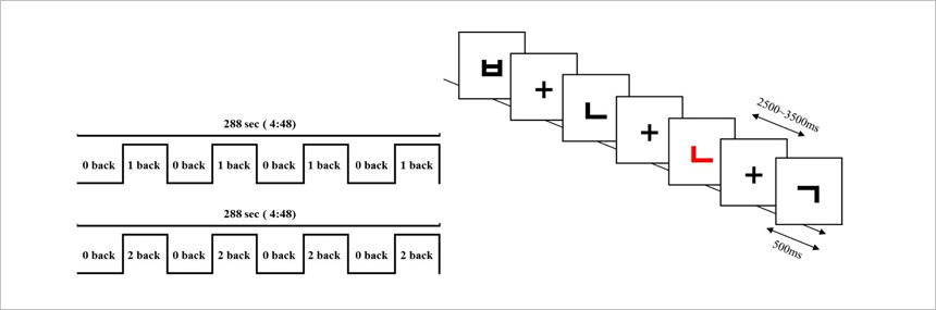
The working memory paradigm consisted of the n-back memory task. The n-back task, where n is an integer (usually 1,2, or 3), requires on-line monitoring, updating, and manipulation of remembered information, and is therefore assumed to place great demands on a number of key processes within working memory.
Participants performed a letter n-back task with two conditions: 0-back and 2-back. In the 0-back condition, participants were asked to remember a larger letter that was presented at the beginning of each trial block. In the 2-back condition, they were asked to respond when a letter matched one that had been presented two letters before the present letter. We used letters from the Korean alphabet as target cues.
2. Wisconsin Card Sorting Task (WCST)
WCST is widely used to examine executive dysfunction. This task requires the participant to match randomly drawn cards to reference cards according to a sorting category of classification of color, shape and number. The sorting category would be changed after a certain number of consecutive correct decisions. Without notice, however, the sorting category is changed and the participant must switch from the previous category to a new sorting category. Thus, the WCST examines a range of cognitive functions including working memory, set shifting, perseveration, and error detection. There is mounting evidence that patients with frontal cortex damage have impaired performance on the WCST.
WCST is consisted of trials with two different test variants (A and B) and a high-level baseline condition (HLB) in an efficient blocked design. Briefly, in task A (РђюuninstructedРђЮ), the stimulus card was preceded by the neutral word РђўcardРђЎ on the screen, giving no information about the sorting criterion. The subject had to determine the sorting criterion by trial and error using each feedback. In task B (РђюinstructedРђЮ), the stimulus card provided information only about a change in the sorting criterion, showing the word РђўshapeРђЎ, РђўcolorРђЎ, or РђўnumberРђЎ on the screen whenever a change occurred. In the HLB condition, subjects had to choose the card identical to one of four reference cards.

Ж▓йвХЂвїђьЋЎЖхљ ВЃЮВ▓┤ВЮўьЋЎВъљЖИ░Ж│хвфЁВўЂВЃЂВЌ░ЖхгВІц(BMRLab) ВъљЖИ░Ж│хвфЁВўЂВЃЂВЮўьЋЎьїђВЌљВёювіћ ВѓгвъїВЮў вЄївЦ╝ ВЌгвЪг ВДђВЌГВю╝вАю вѓўвѕёЖ│а ВЮ┤ ЖхгВЌГВЮ┤ Вќ┤вќц ВЌГьЋаВЮё ьЋўвіћВДђ ВЋїВЋёвѓ┤віћ вЇ░, ВЌгвЪгЖ░ђВДђ в░Ев▓ЋВЮё ВѓгВџЕьЋўВЌг вХёВёЮьЋЕвІѕвІц. Ж░ђВъЦ Ж░ЋваЦьЋю ВаЉЖи╝ в░Ев▓ЋВю╝вАю ВѓгвъївЊцвАю ьЋўВЌгЖИѕ ЖИ░ВќхьЋўЖИ░, ВЮўВѓг Ж▓░ВаЋьЋўЖИ░, вІеВќ┤вЦ╝ ВЃЮЖ░ЂьЋўЖИ░ вЊ▒ВЮў ВёювАю вІцвЦИ ВаЋв│┤вЦ╝ В▓ўвдгьЋўвЈёвАЮ ьЋеВю╝вАюВЇе ЖиИвЊцВЮў вЄїВЌљВёю Вќ┤вќц ВІаЖ▓йьЋЎВаЂ в│ђьЎћЖ░ђ ВЃЮЖИ░віћВДђ ьїїВЋЁьЋўвіћ Ж▓ЃВЮ┤ ВъѕВіхвІѕвІц. Вџ░вдгвіћ вЄї ВІаЖ▓йВёИьЈг ВБ╝в│ђВЮў Вѓ░Вєї вєЇвЈёВЌљ ВЮўВА┤ВаЂВЮИ ВІаьўИвЦ╝ ьєхьЋ┤ ЖИ░віЦВаЂ ВъљЖИ░Ж│хвфЁВўЂВЃЂВЮё ВѓгВџЕьЋўВЌг Вќ┤вќц ВѓгвъїВЮ┤ ьі╣ВаЋ ВаЋв│┤вЦ╝ В▓ўвдгьЋўвіћ Ж│╝ВаЋВЮё ВѕўьќЅьЋўвіћ вЈЎВЋѕ в╣ёВ╣еВіхВаЂВю╝вАю вЄїВЮў вІцВќЉьЋю ВўЂВЌГвЊцВЮё ВаЋВЮўвѓ┤вд┤ Вѕў ВъѕВіхвІѕвІц. Вќ┤вќц ВаЋв│┤ В▓ўвдгВЌљ вїђьЋю taskвЦ╝ ВѕўьќЅьЋўвЕ┤Вёю Вќ┤вќц вЄїВЮў ВўЂВЌГВЮ┤ ьЎюВё▒ьЎћ вљўвіћВДђВЌљ вїђьЋю ВаЋв│┤вЦ╝ ьєхьЋ┤ Вџ░вдгвіћ вЄїЖ░ђ Вќ┤вќ╗Ж▓ї ЖхгВё▒вљўВќ┤ ВъѕвіћВДђвЦ╝ ВЮ┤ьЋ┤ьЋа Вѕў ВъѕВіхвІѕвІц.
TaskВЌљ ЖИ░В┤ѕьЋю ЖИ░віЦВаЂВъљЖИ░Ж│хвфЁВўЂВЃЂВЮў вХёВёЮВЮђ ВѓгвъїВЮў вЄїВЌљВёю ЖИ░віЦВаЂВю╝вАю ЖхгвХё вљўВќ┤ Въѕвіћ ЖхљВаљ(node)вЊцВЮё ЖхгвХёьЋ┤ вѓ┤віћ вЇ░ ВѓгВџЕвљЕвІѕвІц. Ж▓░Ж│╝ВаЂВю╝вАю ЖИ░віЦВаЂ ВъљЖИ░Ж│хвфЁВўЂВЃЂ вХёВёЮВЮђ ью┤ВДђЖИ░ ВЃЂьЃюВЮў ЖИ░віЦВаЂ ВъљЖИ░Ж│хвфЁВўЂВЃЂ, ЖиИвдгЖ│а HARDIвЦ╝ ВЮ┤ВџЕьЋ┤ Вќ╗ВЮђ ВаЋв│┤вАю вЄїВЌљВёю ЖИ░віЦВаЂВю╝вАю ЖхгвХё вљю ЖхљВаљвЊцВЌљ вїђьЋю ВЌ░Ж▓░Вё▒ВЮё ьЋ┤ВёЮьЋўЖ│а ВЮ┤ьЋ┤ьЋўвіћ вЇ░ вЈёВЏђВЮё Вцё Вѕў ВъѕВіхвІѕвІц. Вџ░вдг ВЌ░ЖхгВІцВЮђ Ж░Ђ ьћ╝ьЌўВъљвЊцВЮ┤ ВѕўьќЅьЋўвіћ TaskвЊцВЮё Ж░юв░юьЋўЖ│а ВюаьџеьЋўвЈёвАЮ ВЌ░ЖхгьЋўвЕ░ Ж░юВЮИ в░Ј ЖиИвБ╣ вХёВёЮВЮё Ж░ђвіЦьЋўЖ▓ї ьЋўЖИ░ ВюёьЋ┤ taskвЦ╝ ВёаьЃЮьЋўВЌг ВѓгВџЕьЋўЖ│а ВъѕВіхвІѕвІц.
Taskвіћ Ж░Ђ ьћ╝ьЌўВъљвЊцВЮё В┤гВўЂьЋўвіћ вЇ░ ВєїВџћвљўвіћ ВІюЖ░ё вѓ┤ВЌљВёю ВёювАю вІцвЦИ ВІаЖ▓й ВІюВіцьЁюВЮё ьЈЅЖ░ђьЋа Вѕў ВъѕвЈёвАЮ ЖхгВё▒вљўВќ┤ВЋ╝ ьЋЕвІѕвІц. ВЮ┤вЪгьЋю ЖИ░віЦВаЂВю╝вАю вЄївЦ╝ ЖхгьџЇьЎћьЋўвіћ ВЌГьЋаВЮё ьЋўвіћ taskвЊцВЮђ primary sensoty processes (ВЮ╝В░еЖ░љЖ░Ђ; ВІюЖ░Ђ, Вџ┤вЈЎ ЖИ░віЦ)ВЎђ ВЮИВДђ ЖИ░віЦ, ВаЋВёюВаЂ ЖИ░віЦ, ЖиИвдгЖ│а ВъљЖи╣ВЌљ вїђьЋю в░ўВЮЉ, ВъЉВЌЁ ЖИ░Вќх, ВЮ╝ьЎћВаЂ ЖИ░Вќх, ЖиИвдгЖ│а ВќИВќ┤ ЖИ░віЦ, ВЮўВѓг Ж▓░ВаЋ вЊ▒ВЮё ьЈгьЋеьЋўвіћ вІцВќЉьЋю вЄїВЮў ЖИ░віЦВЮё ьЈЅЖ░ђьЋа Вѕў ВъѕвЈёвАЮ ЖхгВё▒вљўВќ┤ ВъѕВіхвІѕвІц.
1. Working-memory task
ВъЉВЌЁ вЕћвфевдг ьїевЪгвІцВъёВЮђ n-back вЕћвфевдг ВъЉВЌЁВю╝вАю ЖхгВё▒вљЕвІѕвІц. nВЮ┤ ВаЋВѕў (вїђЖ░ю 1, 2 вўљвіћ 3) ВЮИ n-back ВъЉВЌЁВЮђ ЖИ░Вќх вљю ВаЋв│┤ВЮў в╣ёВёаьўЋ вфевІѕьё░вДЂ, ВЌЁвЇ░ВЮ┤ьіИ в░Ј ВА░ВъЉВЮё ьЋёВџћвАю ьЋўЖИ░ вЋївгИВЌљ ВъЉВЌЁ вЕћвфевдг вѓ┤ВЌљВёю ВЌгвЪг ВБ╝Вџћ ьћёвАюВёИВіцВЌљ вїђьЋ┤ ьЂ░ ВЌ░Ж┤ђВё▒ВЮ┤ ВъѕвІцЖ│а Ж░ђВаЋьЋЕвІѕвІц.
В░ИЖ░ђВъљвЊцВЮђ 0-backЖ│╝ 2-backВЮ┤вЮ╝віћ вЉљ Ж░ђВДђ ВА░Ж▒┤ВЮў ЖИђВъљ n-back ВъЉВЌЁВЮё ВѕўьќЅьќѕВіхвІѕвІц. 0-backВЌљВёю В░ИЖ░ђВъљвіћ Ж░Ђ вИћвАЮВЮў ВІюВъЉ вХђвХёВЌљ ВаюВІювљю ьЋюЖИђ ВъљВЮїВЮё ЖИ░ВќхьЋўвЈёвАЮ ВџћВ▓Г в░ЏВЋўВіхвІѕвІц. 2-back ВЃЂьЃюВЌљВёю, ЖиИвЊцВЮђ ьўёВъг в│┤ВЮ┤віћ ЖИђВъљЖ░ђ вЉљ ЖИђВъљ ВаёВЮў ЖИђВъљВЎђ ВЮ╝В╣ў ьЋа вЋї ВЮЉвІхьЋўвЈёвАЮ ВџћВ▓Г в░ЏВЋўВіхвІѕвІц. Вџ░вдгвіћ в│И ВІцьЌўВЌљВёю ьЋюЖИђВЮў ЖИђВъљвЦ╝ ВъљЖи╣Вю╝вАю ВѓгВџЕьќѕВіхвІѕвІц.
2. Wisconsin Card Sorting Task (WCST)
ВюёВіцВйўВІа В╣┤вЊювХёвЦў Ж▓ђВѓгвіћ ВаёВаёвЉљВЌйв░░ВЎИВИА ВўЂВЌГ(Prefrontal Dorsolateral area)ВЮў ЖИ░віЦВъЦВЋавЦ╝ ВИАВаЋьЋўвіћ Ж▓ЃВю╝вАю ВЋївацВДё вїђьЉюВаЂВЮИ ВІцьќЅЖИ░віЦ ьЈЅЖ░ђвЈёЖхгвАюВЇе 'ВѓгЖ│аВЮў ВюаВЌ░Вё▒(Flexibility in thinking)' ьў╣ВЮђ ВЮИВДђВаЂ ВюаВЌ░Вё▒(Cognitive Flexibility)'ВЮё ВИАВаЋьЋўЖИ░ ВюёьЋ┤ Ж░юв░ювљўВЌѕВіхвІѕвІц.
ВЮ┤ьЏё В╣┤вЊювХёвЦўЖ▓ђВѓгВЌљвіћ ВХћВЃЂВаЂ Ж░ювЁљ ьўЋВё▒(Concept generation)ВЮё ьЋўЖ│а в▓ћВБ╝ьЎћ ьЋўвіћ віЦваЦ, ВЮИВДђВаЂ вЈёВІЮВЮў в│ђьЎћ(Cognitive set shifting), ВЌгвЪгЖ░юВЮў ВъљЖи╣ ВцЉ Ж┤ђваеВЌєвіћ ВъљЖи╣ВЌљ вїђьЋю в░ўВЮЉВЮё ВќхВаюьЋўЖ│а ВЮ┤в»И ВёаьЃЮьЋю в░ўВЮЉ ВЏљВ╣ЎВЌљ вћ░вЮ╝ в░ўВЮЉьЋўвацвіћ Ж▓йьќЦВЮё ВќхВаюьЋўвацвіћ віЦваЦ(The ability to inhibit prepotent responses), ВІюЖ░ЂВаЂВю╝вАю ВёИвХђВаЂВЮИ ьі╣ВДЋВЮё в│ђв│ёьЋўвіћ віЦваЦ(Attribute identification). Ж░ђВёцВЮё ВёцВаЋьЋўЖ│а вгИВаювЦ╝ ьЋ┤Ж▓░ьЋўЖ▒░вѓў ВХћвдгьЋўвіћ віЦваЦ, Ж│ёьџЇ в░Ј ВА░ВДЂьЎћ віЦваЦ, ЖиИвдгЖ│а ВДђВєЇВаЂВЮИ ВБ╝ВЮўваЦВЮ┤ ьЋёВџћьЋю Ж▓ЃВю╝вАю ВЋївацВаИ ВъѕВіхвІѕвІц.
ВЮ┤ В╣┤вЊю вХёвЦў Ж▓ђВѓгВЌљВёювіћ В░ИЖ░ђВъљЖ░ђ ВЃЅВЃЂ, вфеВќЉ в░Ј ВѕФВъљВЮў вХёвЦў ЖИ░ВцђВЌљ вћ░вЮ╝ вѓўВЌ┤вљю В╣┤вЊювЦ╝ В░ИВА░ В╣┤вЊюВЎђ вїђВА░ьЋ┤ВЋ╝ ьЋЕвІѕвІц. вХёвЦў ЖИ░ВцђВЮђ ВЌ░ВєЇВаЂВЮИ Вўгв░ћвЦИ Ж▓░ВаЋ ВЮ┤ьЏёВЌљ в│ђЖ▓йвљўвѓў ВЋёвг┤вЪ░ ВДђВІю ВЌєВЮ┤ вХёвЦў ЖИ░ВцђВЮђ в│ђЖ▓йвљўЖ│а В░ИЖ░ђВъљвіћ ЖИ░ВА┤ВЮў ЖИ░ВцђВЌљВёю ВЃѕвАюВџ┤ ЖИ░ВцђВю╝вАю в╣авЦ┤Ж▓ї ВЮИВДђвЦ╝ ВаёьЎўьЋ┤ВЋ╝ ьЋЕвІѕвІц.
![]()
601, 515-3, Hakjeong-dong, Buk-gu, Daegu 702-911, Republic of KOREA | Tel : +82-53-950-4128, 4129 | e-mail : ychang@knu.ac.kr
Copyright(c) 2016 Kyungpook National University School of Medicine BMR lab. All rights reserved.